Ophthalmology
The reference book of Ophthalmology

Q: How many ophthalmic medical conditions are there?
A: There are more than 354 ophthalmic medical disorders.
Here are further guidelines.

What are the skills and knowledge required of an ophthalmologist?
  1. Eye emergencies

  2. Common, non-emergency eye problems

  3. Consultation documentation

  4. Research documentation

  5. Administrative issues

  6. Teaching skills relevant to this profession, preferably via the Internet

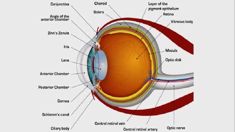
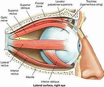
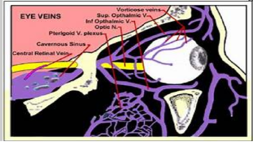
Anatomy, Physiology and Pathology of the Human Eye
  1. Anterior chamber

  2. Bulba sheath

  3. Canal of Schlemm

  4. Central retinal artery

  5. Central retinal vein

  6. Choroid

  7. Ciliary muscle

  8. Ciliary process

  9. Ciliary zonules

  10. Conjunctiva

  11. Cornea

  12. Dura mater

  13. Fovea centralis

  14. Inferior oblique muscle

  15. Inferior rectus muscle

  16. Iris

  17. Lenticular cortex

  18. Lenticular nucleus

  19. Macula lutea

  20. Medial rectus muscle

  21. Optic disc

  22. Optic nerve

  23. Ora serrata

  24. Posterior compartment

  25. Pupil

  26. Retina

  27. Retinal arteries / veins

  28. Sclera

  29. Superior rectus muscle

  30. Vorticose vein

Eye Emergencies
Eye Emergencies: Types, Symptoms, and Prevention

Ophthalmologist
Sudden Loss of Vision
Sudden Loss of Vision and Slow Loss of Vision
  1. What are the symptoms of an eye emergency? Answer

  2. What are the causes of a sudden painful loss of vision? Answer

  3. What are the causes of a sudden painless loss of vision? Answer

  4. What are the causes of a gradual painful loss of vision? Answer

  5. What are the causes of a gradual painless loss of vision? Answer

  6. Have you asked about the proper medical history relevant to this case? Answer

  7. What are the 8-point eye exam findings according to the American Academy of Ophthalmology? Answer

  8. What is the treatment? Answer

Eye Emergency Symptoms
What are symptoms of an eye emergency?
1. Sudden loss of vision in both eyes/sudden blindness
2. Sudden loss of vision in the left eye
3. Sudden loss of vision in the right eye
4. Bleeding from the eye
5. Blood in the white part of the eye
6. Bruising around the eye
7. Burning or stinging
8. Decreased vision
9. Discharge from the eye
10. Double vision
11. Eye pain
12. Eye trauma: penetrating and/or perforating injury
13. Eye trauma: non-perforating
14. Light sensitivity
15. New or severe headaches
16. One eye is not moving like the other
17. One eye is sticking out or bulging
18. Pupils that are not the same size
19. Redness and irritation
20. Severe itching
What are the causes of sudden painful loss of vision?
Sudden painful loss of vision can be due to the following causes:
(1) Keratitis
(2) Conjunctivitis
(3) Scleritis
(4) Uveitis
(5) Angle closure glaucoma

What are the causes of sudden painless loss of vision?
Sudden painless loss of vision can be due to the following causes:
(1) Retinal vascular occlusion
(2) Retinal detachment
(3) Optic neuritis
(4) Retinoblastoma

What are the causes of gradual painful loss of vision?
Gradual painful vision loss can be due to the following causes: (1) Keratitis (2) Scleritis (3) Chronic iridocyclitis

What are the causes of gradual painless loss of vision?
Gradual painless loss of vision can be due to the following causes:
(1) Cataract
(2) Refractive error
(3) Diabetic retinopathy
(4) Age-related macular dystrophy/macular degeneration (painless)
(5) Retinitis pigmentosa

Blindness

Blindness: What is it?
Being unable to see from both eyes.

What is another word or term for being blind?
Being completely blind

Is there a difference between being blind and being visually impaired?
Yes.

What is vision loss/visual impairment?
Vision loss is a state of impairment to the vision of the eye, which can be complete or partial, permanent or temporary, and unilateral or bilateral.

Sudden loss of vision and slow loss of vision: What is the difference?
The causes are different.

How many causes of sudden loss of vision are there?
At least 11.

What are the causes of sudden loss of vision?
1. Detachment of the retina
2. Occlusion of the retinal vessel or artery
3. Vitreous hemorrhage
4. Amaurosis fugax
5. Glaucoma
6. Optic neuritis
7. Ocular migraine
8. Drugs
9. Giant cell arteritis
10. Trauma
11. Other conditions

Medical history relevant to the issue

Have you asked about the proper medical history relevant to this case?

What is the medical history relevant to this case?

How can visual impairment be described?

Complete
Partial
Permanent
Temporary
Unilateral
Bilateral
Sudden
Gradual
Painful
Painless
The person can see normally relevant to their age.
The person has come in for a routine checkup.

How is visual loss classified?
Sudden loss of vision
Gradual loss of vision
Painful loss of vision
Painless loss of vision

What medical history is necessary for sudden loss of vision?

When did it start?

Where did it start?

How did it start?

How long has this been present?

Was this sudden or has this progressed?

Did the sudden loss of vision occur with pain or without pain?

Does the vision loss affect one or both eyes?

Is the vision loss total or does it affect only a specific part of the field of vision?

Are there other vision symptoms, such as floaters, flashing lights, halos around lights, distorted color vision, jagged or mosaic patterns, and/or eye pain?

Are there any risk factors that may cause eye problems?


What are the 8-point eye exam findings according to the American Academy of Ophthalmology?
1. Visual acuity
2. Pupils
3. Extraocular motility and alignment
4. Intraocular pressure
5. Confrontation visual fields
6. External examination
7. Slit-lamp examination
8. Fundoscopic examination

What is the treatment?
Treat the underlying cause.
Term Definition
Abrasion Defect of the corneal epithelium. Stains with fluorescein. Usually heals within 24 – 48 hours
Contusion Result of blunt injury, either at site of impact or distant to it
Closed injury The wall of the globe is intact but structures inside the eye are damaged
Rupture Jagged open injury due to blunt injury, often away from the site of injury at the weakest points of the globe: concentric to the limbus, just behind the insertion of the extraocular muscles or at the equator
Open injury A full thickness break in the wall of the eye; can follow either sharp or severe blunt injury
Lamellar laceration Partial thickness wound caused by a sharp object
Laceration Full thickness penetration
Penetration Entry wound only
Perforation ‘Through and through’ injury; an injury that goes right through the eye, causing both entry and exit wounds
Emergency Care
If you have a problem that you feel is an emergency or life threatening, call 911 immediately or go to the nearest emergency room.

What are ophthalmological medical emergencies?
How should you do a quick assessment, diagnosis, and treatment of a person reported as an ophthalmological medical emergency?


* Acute Angle-Closure Glaucoma
* Acute Multifocal Placoid Pigment Epitheliopathy
* Acute Orbital Compartment Syndrome
* Acute Retinal Necrosis
* Alacrima
* Angle Recession Glaucoma
* Aphakic and Pseudophakic Glaucoma
* Best Disease
* Central Serous Chorioretinopathy
* Chalazion in Emergency Medicine
* Complications and Management of Glaucoma Filtering
* Conductive Keratoplasty Hyperopia and Presbyopia
* Congenital Anomalies of the Nasolacrimal Duct
* Corneal Laceration
* Corneal Ulceration and Ulcerative Keratitis in Emergency Medicine
* Dacryoadenitis
* Dacryocystitis
* Drug-Induced Glaucoma
* Dry Eye Syndrome
* Eales Disease
* Emergency Care of Corneal Abrasion
* Emergent Treatment of Acute Conjunctivitis
* Endophthalmitis
* Exudative Retinal Detachment
* Fuchs Heterochromic Uveitis
* Glaucoma, Angle Closure, Acute
* Glaucoma, Angle Closure, Chronic
* Glaucoma, Suspect, Adult
* Globe Rupture
* Hordeolum and Stye in Emergency Medicine
* Hyphema Glaucoma
* Intermediate Uveitis
* Intraocular Foreign Body
* Iris Prolapse
* Iritis and Uveitis
* Juvenile Glaucoma
* Juvenile Idiopathic Arthritis Uveitis
* Lacrimal Gland Tumors
* LASIK Hyperopia
* Lattice Degeneration
* Lens-Particle Glaucoma
* Low-Tension Glaucoma
* Macular Hole
* Nonpseudophakic Cystoid Macular Edema
* Obstruction Nasolacrimal Duct
* Ocular Burns
* Ocular Hypotony
* Periorbital Infections
* Phacolytic Glaucoma
* Phacomorphic Glaucoma
* Phakic IOL Hyperopia
* Pigmentary Glaucoma
* Plateau Iris Glaucoma
* Posner-Schlossman Syndrome
* Postoperative Corneal Edema
* Postoperative Endophthalmitis
* Primary Congenital Glaucoma
* Primary Open-Angle Glaucoma
* Proliferative Retinal Detachment
* Pseudoexfoliation Glaucoma
* Retinal Artery Occlusion
* Retinal Detachment
* Retinal Vein Occlusion
* Rhegmatogenous Retinal Detachment
* Secondary Congenital Glaucoma
* Sudden Visual Loss
* Toxic Anterior Segment Syndrome
* Tractional Retinal Detachment
* Ultraviolet Keratitis
* Unilateral Glaucoma
* Uveitic Glaucoma
* Uveitis, Anterior, Childhood
* Uveitis, Anterior, Granulomatous
* Uveitis, Anterior, Nongranulomatous
* Vitreous Hemorrhage in Emergency Medicine
Eye Trauma

Managing Serious Cases of Ocular Trauma

Assessing and managing eye injuries

Definitions of terms used to describe eye injuries

Eye trauma refers to damage caused by a direct blow to the eye. The trauma may affect not only the eye, but the surrounding area, including adjacent tissue and bone structure.

There are many different forms of trauma, varying in severity from minor injury to medical emergencies. Even in cases where trauma seems minor, every eye injury should be given medical attention.

What Causes Eye Trauma?

When the eye is hit with blunt force, it suddenly compresses and retracts. This can cause blood to collect underneath the hit area, which leads to many of the common symptoms of eye trauma.

Symptoms of Eye Trauma

Symptoms of eye trauma may include:
•Pain
•Trouble seeing
•Cuts to the eyelid
•One eye not moving as well
•One eye sticks out
•Blood in the clear part of the eye
•Unusual pupil size or shape
•Something embedded in the eye
•Something under the eyelid that cannot be easily removed

Treatments for Eye Trauma

Every eye injury should be given medical attention; do not touch, rub or try to remove any object in the eye. If the eye has been cut or there is an object in the eye, rest a protective shield – such as a paper cup – on the bone around your eye. Make sure there is no pressure on the eye itself. Seek immediate, professional medical attention.

In minor cases of trauma, such as a black eye from a sports injury, applying cold to the affected area can help bring swelling down, and allow the affected area to heal faster. However, even in cases where trauma seems minor, every eye injury should be given medical attention.

The best way to avoid eye trauma is to prevent it by using protective eyewear while doing things that may put them at risk. Activities include home repair, yard work, cleaning, cooking, and playing sports. In most cases of injury, people report not properly protecting their eyes – which shows that proper precautions may prevent an eye injury.

Most common eye conditions
Age-Related Macular Degeneration (AMD)
Amblyopia (Lazy Eye)
Astigmatism
Cataracts
Corneal Disease
Diabetic Retinopathy
Dry Eye
Eye Disease Simulations
Floaters
Glaucoma
Hyperopia (Farsightedness)
Low Vision
Myopia (Nearsightedness)
Presbyopia
Refractive Errors
Retinopathy of Prematurity (ROP)
Retinal Detachment
Uveitis

Eye Care
Common, Non-Emergency Eye Problems
Are you having any problems with your vision?
How long have you had these problems?
When do these problems occur?
When was your last eye examination?
Do you have any family history of eye problems?
How is your general health?
What medications are you taking?
Do you have any allergies?
Do you wear glasses/contacts now?
Have your glasses/contacts become stronger over the years?
Do you work with a computer?
Do your eyes ever tire (burn, itch) when reading?
Do you ever see double images or halos around images?
Do you suffer from eye strain and/or tension headaches?
Here are further guidelines.
http://www.qureshiuniversity.com/eyecare.html
Ophthalmologist

What is an ophthalmologist?
How is an ophthalmologist different from an optometrist and an optician?
What is a subspecialist?
When should I see an ophthalmologist?
What happens during an eye examination?
What treatments are available for my eyes?
Here are further guidelines.
Q: What should you know and be able to do as an ophthalmologist?

What is an ophthalmologist?
You are first a physician, than a specialist or super specialist.
Ophthalmology is the study of medical conditions relating to the eye. Ophthalmologists are doctors who specialize in the medical and surgical treatment of this organ. A general practice doctor may refer someone to an ophthalmologist if they show symptoms of cataracts, eye infections, optic nerve problems, or other eye conditions.

An ophthalmologist is a medical doctor who specializes in diagnosing and treating eye-related conditions.

Some ophthalmologists undergo a year or two of fellowship training specializing in one of the many subspecialties of ophthalmology, such as:
glaucoma
the cornea
the retina
uveitis
refractive surgery
pediatrics
neuro-ophthalmology
plastic and reconstructive surgery
ocular oncology
Subspecialist ophthalmologists have usually completed training that allows them to work on eye conditions that are complex, involve a specific part of the eye, or affect certain groups of people. They also train more extensively than regular ophthalmologists to perform extremely intricate surgeries on delicate parts of the eye.

What conditions do they treat?
Ophthalmologists are responsible for the diagnosis, prevention, and treatment of almost all eye conditions and visual issues.

However, subspecialist ophthalmologists tend to treat and monitor certain conditions, such as:

glaucoma
retinal conditions, such as macular degeneration and diabetic retinopathy
corneal conditions
cataracts
cases involving children or childhood eye conditions
cases with a neurological cause or component, such as optic nerve problems, abnormal eye movements, double vision, and some kinds of vision loss
cases involving complex surgical procedures, such as reconstructive surgery or advanced vision repair
Aside from caring for the eyes and vision, an ophthalmologist’s medical training may also equip them to notice symptoms of conditions that do not directly relate to the eye. In such cases, they can refer people for the appropriate treatment.

Many ophthalmologists also participate in some form of scientific research focusing on the causes of eye and vision conditions, as well as potential cures.

What procedures do they do?
Most ophthalmologists are trained and certified to perform a wide range of medical and surgical procedures. The procedures that an ophthalmologist regularly carries out depend on several factors, such as the type of practice and specialty in which they work.

Some of the most common everyday procedures that an ophthalmologist will perform include diagnosing and monitoring mild eye and vision conditions. They will also spend time prescribing and fitting glasses and contact lenses to correct vision problems.

Subspecialist ophthalmologists tend to perform a smaller range of procedures on a day-to-day basis, focusing instead on the treatment of one condition or a few related conditions.

Procedures that subspecialists commonly perform include:

  • diagnosis and monitoring of moderate-to-severe eye conditions
  • cataract surgery
  • glaucoma surgery
  • refractive surgery to correct vision
  • cancer treatment
  • reconstructive surgery to repair trauma or birth abnormalities, such as crossed eyes
  • chronic or severe tear duct infections or blockages
  • neoplasm (tumor, cyst, or foreign object) removal
  • monitoring or consulting on cases relating to other conditions, such as diabetic retinopathy or immune conditions
  • injections around the eyes and face to alter facial structure function and appearance
  • repairing torn or detached retinas
  • corneal transplants
When to see an ophthalmologist

Most people see an ophthalmologist because they are experiencing chronic or severe vision symptoms or signs of eye conditions, such as:

  • bulging eyes
  • reduced, distorted, blocked, or double vision
  • excessive tearing
  • eyelid abnormalities or problems
  • seeing colored circles or halos around lights
  • misaligned eyes
  • black specks or strings called floaters in the field of view
  • seeing flashes of light
  • unexplained eye redness
  • loss of peripheral vision

A person may need emergency care from an ophthalmologist if their symptoms include:

  • sudden vision loss or changes
  • sudden or severe eye pain
  • eye injury

A person may also receive a referral to an ophthalmologist if they have conditions or factors that can increase the risk of eye conditions, such as:

  • high blood pressure
  • diabetes
  • a family history of eye conditions
  • thyroid conditions, for example, Graves’ disease

A family doctor, pediatrician, emergency room doctor, or optometrist usually refers a person to an ophthalmologist.

The American Academy of Ophthalmology recommend that people have a full medical eye exam by the age of 40 years so that an ophthalmologist can create a baseline profile of their eye health.

Having an eye health baseline is important because it makes it easier for doctors to spot or track eye or vision changes, which are often subtle and difficult to detect. Even healthy people can suddenly experience severe eye conditions.

Vision tests and eye exams are procedures an optometrist may perform.

Unlike ophthalmologists, optometrists and opticians are not medical doctors. However, members of all three distinct professions can, and frequently do, work in the same office or practice.

Optometrists are healthcare professionals who provide primary vision care. Optometrists hold a Doctor of Optometry (OD) degree, which requires the completion of 3–4 years of college and then 4 years of optometry school.

While the procedures that they perform vary between states and individual practices or clinics, most optometrists:

  • perform vision tests and eye exams
  • prescribe and dispense corrective lenses
  • help manage and monitor vision changes
  • detect signs of conditions that need subspecialist care, such as glaucoma and cataracts
  • prescribe medications to help manage certain conditions

Opticians are a type of healthcare technician. They are specially trained to help design, confirm, select, or fit corrective vision devices, including contact lenses and eyeglass lenses and frames. Opticians cannot diagnose or treat conditions and must follow the prescription and guidance of optometrists and ophthalmologists.

The other eye healthcare professionals who frequently work with ophthalmologists and optometrists include:

  • ophthalmic medical assistants: these technicians perform many different tests and assist ophthalmologists
  • ophthalmic technicians: these more highly trained or experienced medical assistants help ophthalmologists perform more complex tests and minor office surgeries
  • ophthalmic photographer: these professionals use special cameras and photography methods to create pictures of the eyes that help document eye conditions
  • ophthalmic registered nurse: these healthcare clinicians have specialized nursing training and can help ophthalmologists perform technical tasks, such as assisting with surgeries or injecting medications
Pediatric Ophthalmology

Q: How do you diagnose and treat ophthalmic medical conditions in children?

Eye Testing for Newborn
    How do you do an eye exam of a newborn?
    Who should do an eye exam of a newborn?
    What should you look for in an eye exam of a newborn?
Eye Testing for Infants
    Q: How do you do an eye exam for a six month-old?
Eye Testing for Preschool Children
    Q: Does your child seem to see well?
    Q: Does your child hold objects close to his or her face when trying to focus?
    Q: Do your child's eyes appear straight or do they seem to cross or drift or seem lazy?
    Q: Do your child's eyes appear unusual?
    Q: Do your child's eyelids droop or does 1 eyelid tend to close?
    Q: Have your child's eye(s) ever been injured?
Ophthalmology History
What Does 20/20 Vision Mean?
Glossary of Ophthalmology Terms
Ophthalmology Sections
Snellen Eye Test
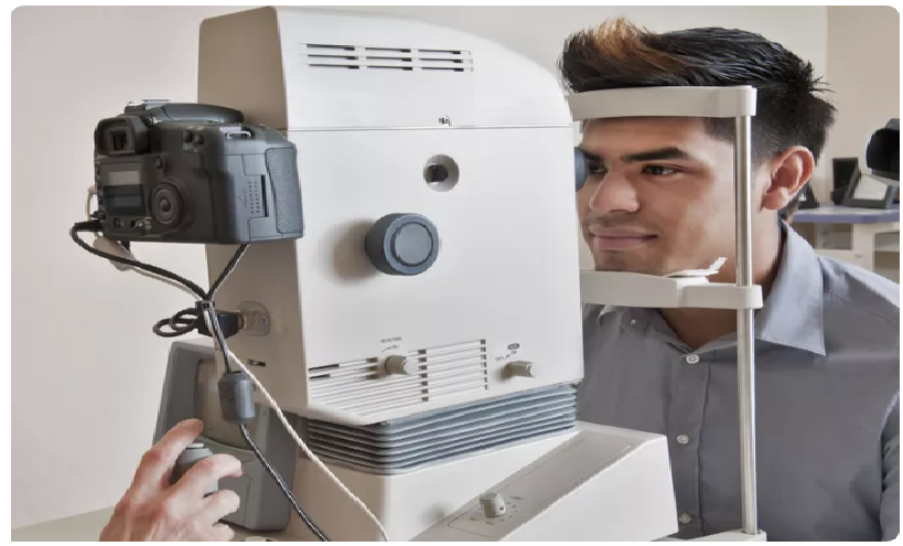
Digital Retinal Photography
Corneal Topography
Computerized Automated Refractor
    What is a computerized automated refractor?
    Who are the manufacturers of automated refractors?
    What are the uses of a computerized automated refractor?
    How is a computerized automated refractor test administered?
    What are common types of glasses/spectacles?
Automated Non-contact Tonometer
Automated Visual Field Perimetry
Marco TRS Automated Refraction System
Computerized Patient Records System
Ophthalmic instrument and equipment manufacturers
    What are various ophthalmic instruments and equipment?
    Who are the quality ophthalmic instrument and equipment manufacturers?
    What materials do you need to manufacture ophthalmic instruments and equipments?
    How are ophthalmic instruments and equipments manufactured?
    Here are further guidelines.
Refractive errors
    Q: What is normal vision?
    Q: What does it mean when someone has 20/20 vision?
    Q: What are refractive errors?
    Q: What causes refractive errors?
    Q: What are the symptoms?
    Q: What is the difference between reading glasses and distance glasses?
    Q: How often and how long should plus lenses be used?
    Q: What if problems such as tired eyes are experienced when using plus lenses?
    Q: Why do many people need stronger minus glasses year after year?
    Q: Are plus lenses harmful in any way?
    Q: How far should the eyes be from the book or computer or other close object when using the reading glasses?

    Spectacles
    Fashionable Frames
    Quality Lenses

    Q: What are Polarised lenses?
    Q: What are Transition lenses?
    Q: What are Polycarbonate lenses?
Eye Injuries
    Q: What are the symptoms of eye injuries?
    Q: How are eye injuries diagnosed?
    Q: How are eye injuries treated?
Low Vision
Eye Safety
Retinal Detachment
Cataract
    Eye Cataract is quite common especially in the old age. What are the reasons responsible for this condition?
    What is the best treatment available for treating cataract?
Diabetic retinopathy
    How does Diabetes affect the eyes and what is the treatment?
Health care and desktop computer monitor
Laser vision correction surgery
Retina and vitreous consultation including evaluation and treatment of age related macular degeneration
Cornea transplantation
Management and treatment of eyelid and orbital cancers
Management and treatment of thyroid eye disease
Cosmetic eyelid surgery
Reconstructive eyelid and orbital surgery
Glaucoma evaluation and treatment
Evaluation of all pediatric eye conditions
Evaluation of genetic conditions which affect the eye or visual system
Eye Health Organizations
People often complain of red eyes, frequent watering, and eye strain. What precautions should people take to avoid it?
Computer vision syndrome is getting common these days with the increased use of computers.
What is the solution?
What are the precautions you would advise for general eye care?

What is the difference between an ophthalmologist, an optometrist and an optician?
What are the best things about your specialty?
What are the worst things about your specialty?
Why did you choose your specialty?
What types of clinical cases do you commonly see?
Briefly describe a typical day.
What are the varieties of lifestyles within your field?
How do you see your discipline changing over the next decade?
On November 14, 2010, an 18-year-old boy from Baramullah was diagnosed with trauma and retinal detachment at SKIMS. Is that correct?
Treatment outside the state was advised.
Why does a retinal detachment need treatment outside the state?
How many ophthalmologists are there in the state?
Why didn't the state department of health impart training to existing medical doctors or ophthalmologists to treat such cases within the state?
Eye Disability
    When do you declare that a person has eye disability?
There is no exact verified number of blind people in the state.
There is no exact verified number of people with eye disabilities in the state.
A Community Ophthalmology Unit is opened at a primary health center.
How did they determine that a Community Ophthalmology Unit at a primary health center at specific location is required?
Who has the duty to maintain an exact verified number of blind people and those with eye disabilities in the state?
Who has the duty to research and treat these cases?

How many people in your state are completely blind?
What is the cause of blindness?
What is their profile?
Where is the exact lesion?
Is their a hope for them to regain their vision?
What have you done?
How can I/we help?

Are you as an ophthalmologist facing a most difficult case?
If yes, feel free to communicate and consult.

admin@qureshiuniversity.com

Ophthalmic instrument and equipment manufacturers
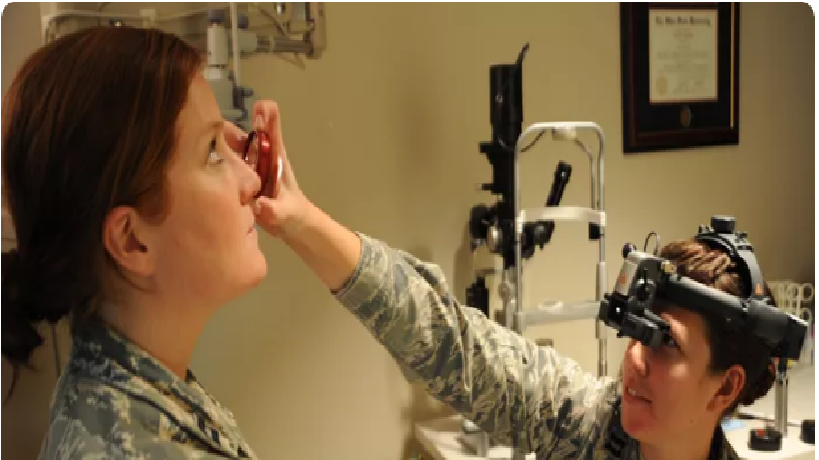
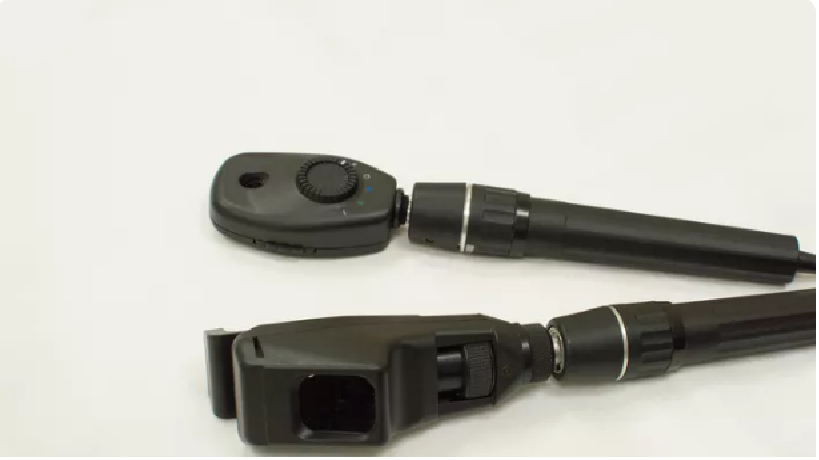
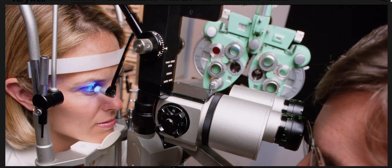
Gallery of Eye Examination Equipment.
What are various ophthalmic instruments and equipment? Answer
How brightly lit is an eye exam room?
What are "normal" results of an eye exam?
What should be the luminous intensity of a desktop computer monitor?
What will you advise as a medical doctor or ophthalmologist to a patient using a desktop computer monitor for a prolonged time?
Who are the quality ophthalmic instrument and equipment manufacturers?
What materials do you need to manufacture ophthalmic instruments and equipments?
How are ophthalmic instruments and equipments manufactured?

What are the consequences of higher luminous intensity of a desktop computer monitor?
People take a prolonged time to become accustomed to normal room luminous intensity after prolonged activities on the desktop computer monitor.

How could this be prevented?
Brightness/contrast of the desktop computer monitor
60/31
This needs ongoing research.

Here are further guidelines.

What are various ophthalmic instruments and equipment?
1.
Autorefractor
2. Binocular Indirect Ophthalmoscope
3. Lensometer
4. Manual Keratometer
5. Phoropter
6. Retinal Camera
7. Retinoscope and Direct Ophthalmoscope
8. Slit Lamp
9. Tonometer
10. Ophthalmology Operating Microscopes | Cataract Surgery Equipment
11. List of instruments used in ophthalmology
12. Eye Exam
Ophthalmology Clinic Design Guide for Optometric clinic


Autorefractor

An autorefractor is a machine used to measure a person's refractive error and prescription for eyeglasses or contact lenses. This is achieved by measuring how light is changed as it enters a person's eye.

The automated refraction technique is quick, simple and painless. The patient takes a seat and places their chin on a rest. One eye at a time, they look into the machine at a picture inside. The picture moves in and out of focus as the machine takes readings to determine when the image is on the retina.

Several readings are taken which the machine averages to form a prescription. No feedback is required from the patient during this process.
Binocular Indirect Ophthalmoscope

An ophthalmoscope is an instrument used for examining the interior structures of the eye, especially the retina, consisting of a mirror that reflects light into the eye and a central hole through which the eye is examined.

A binocular indirect ophthalmoscope (BIO) is worn on an eye doctor's head in order to have the use of both hands to examine the eyes.
Lensometer

A lensometer is an instrument used to measure the power of an existing lens. An optician uses a lensometer to determine the prescription of a patient's eyeglasses.
Manual Keratometer

A manual keratometer is used to determine how flat or steep the cornea is. It is often used to measure and diagnose conditions such as astigmatism, keratoconus, corneal scarring and corneal distortion. A keratometer is commonly used to fit contact lenses as well.
Phoropter

A phoropter (or phoroptor) is an instrument used during an eye examination to measure refractive error and determine eyeglass prescriptions. Typically, the patient sits behind the phoropter and looks through it at an eye chart.

The optometrist then changes lenses and other settings, while asking the patient for feedback on which settings give the best vision.
Retinal Camera

A retinal camera is used to photograph the back of the eye, including the retina. It is used to document eye diseases. The camera produces a bright flash when a picture is taken.
Retinoscope and Direct Ophthalmoscope

A retinoscope is used to shine light into a patient's eye for an eye doctor to observe the reflection off the retina. The light is moved back and forth across the pupil

A retinoscope is especially useful in prescribing corrective lenses for patients who are unable to give oral feedback to the eye doctor. It is also useful for determining how well the eyes work together (accommodate) to see clearly.

A direct ophthalmoscope is a hand-held instrument used for examining the interior structures of the eye, especially the retina. It consists of a mirror that reflects light into the eye and a central hole through which the eye is examined.
Slit Lamp

A slit lamp is a microscope with a light attached that allows the doctor to closely examine the eye. This instrument is used to view the structures of the eye such as the cornea, iris, and lens.

With special lenses, it is possible to examine the back of the eye as well. A slit lamp allows the practitioner to have an amazing view of the inside of your eyes.

Tonometer

A tonometer is used to measure the pressure of the eye. The test is used to help detect glaucoma. Numbing drops are used for the type of tonometer that actually touches the eye. Some doctors use the air-puff tonometer in which no numbing drops are needed.

A tonometer measures the production of aqueous humor, the liquid found inside the eye, and the rate at which it drains into the tissue surrounding the cornea.
Ophthalmology Operating Microscopes | Cataract Surgery Equipment


Carl Zeiss is the first to develop ophthalmology operating microscopes with Stereo Coaxial Illumination (SCIâ„¢). With this technology, you and your patients will benefit from:

Unique detail recognition
High-contrast brilliance and stability of the red reflex
Even in patients with strongly pigmented, decentered and ametropic eyes, this cataract surgery equipment technology enables you to easily see all the eye’s details.

Eye Diseases and Disorders
Q: How many ophthalmic medical conditions are there?
A: There are more than 350 ophthalmic medical disorders.

What are various examples?
  1. Age-Related Macular Degeneration (AMD)

    Ophthalmology Sections
    Anterior Chamber

  2. Hyphema

  3. Postoperative Flat Anterior Chamber

  4. Synechia, Peripheral Anterior

  5. Toxic Anterior Segment Syndrome
    Choroid

  6. Angioid Streaks

  7. Choroidal Detachment

  8. Choroidal Rupture

  9. Melanoma, Choroidal

  10. Multifocal Choroidopathy Syndromes

  11. Neovascularization, Choroidal
    Conjunctiva

  12. Cicatricial Pemphigoid

  13. Conjunctivitis, Allergic

  14. Conjunctivitis, Bacterial

  15. Conjunctivitis, Giant Papillary

  16. Conjunctivitis, Neonatal

  17. Conjunctivitis, Viral

  18. Filtering Bleb Complications

  19. Keratoconjunctivitis, Epidemic

  20. Melanoma, Conjunctival

  21. Papilloma, Conjunctival

  22. Pharyngoconjunctival Fever

  23. Pterygium

  24. Squamous Cell Carcinoma, Conjunctival

  25. Subconjunctival Hemorrhage
    Connective Tissue Disorders

  26. Ankylosing Spondylitis

  27. Pseudoxanthoma Elasticum

  28. Reactive Arthritis

  29. Sjogren Syndrome
    Cornea

  30. Central Sterile Corneal Ulceration

  31. Congenital Clouding of the Cornea

  32. Contact Lens Complications

  33. Corneal Abrasion

  34. Corneal Edema, Postoperative

  35. Corneal Erosion, Recurrent

  36. Corneal Foreign Body

  37. Corneal Graft Rejection

  38. Corneal Melt, Postoperative

  39. Corneal Mucous Plaques

  40. Dermoid, Limbal

  41. Descemet Membrane Folds

  42. Dystrophy, Crystalline

  43. Dystrophy, Fuchs Endothelial

  44. Dystrophy, Granular

  45. Dystrophy, Lattice

  46. Dystrophy, Macular

  47. Dystrophy, Map-dot-fingerprint

  48. Herpes Simplex

  49. Keratitis, Bacterial

  50. Keratitis, Fungal

  51. Keratitis, Herpes Simplex

  52. Keratitis, Interstitial

  53. Keratoconjunctivitis, Atopic

  54. Keratoconjunctivitis, Sicca

  55. Keratoconjunctivitis, Superior Limbic

  56. Keratoconus

  57. Keratopathy, Band

  58. Keratopathy, Neurotrophic

  59. Keratopathy, Pseudophakic Bullous

  60. Laceration, Corneoscleral

  61. Megalocornea

  62. Neovascularization, Corneal, CL-related

  63. Pellucid Marginal Degeneration

  64. Peripheral Ulcerative Keratitis

  65. Posterior Polymorphous Corneal Dystrophy

  66. Thygeson Superficial Punctate Keratitis

  67. Ulcer, Corneal
    Computer Vision Syndrome

  68. Computer Vision Syndrome

    Dermatologic Disorders

  69. Acrodermatitis Enteropathica

  70. Dermatitis, Atopic

  71. Dermatitis, Contact

  72. Ichthyosis

  73. Kaposi Sarcoma

  74. Kawasaki Disease

  75. Ocular Rosacea

  76. Psoriasis

  77. Stevens-Johnson Syndrome
    Endocrine Disorders

  78. Pituitary Apoplexy
    Extraocular Muscles

  79. Abducens Nerve Palsy

  80. Brown Syndrome

  81. Convergence Insufficiency

  82. Duane Syndrome

  83. Esotropia and Exotropia, A-patterns

  84. Esotropia and Exotropia, V-patterns

  85. Esotropia, Accommodative

  86. Esotropia, Acquired

  87. Esotropia, Infantile

  88. Esotropia, Pseudo

  89. Esotropia, with High AC/A Ratio

  90. Exotropia, Acquired

  91. Exotropia, Congenital

  92. Exotropia, Pseudo

  93. Globe Retraction

  94. Hermansky-Pudlak Syndrome

  95. Monofixation Syndrome

  96. Nystagmus, Acquired

  97. Nystagmus, Congenital

  98. Oculomotor Nerve Palsy

  99. Trochlear Nerve Palsy
    Genetic Disorders

  100. Albinism

  101. Aniridia in the Newborn

  102. Down Syndrome

  103. Familial Dysautonomia

  104. HLA-B27 Syndromes

  105. Peters Anomaly

  106. Prenatal Diagnosis for Congenital Malformations and Genetic Disorders
    Globe

  107. Anophthalmos

  108. Endophthalmitis, Bacterial

  109. Endophthalmitis, Postoperative
    Hematologic and Cardiovascular Disorders

  110. Giant Cell Arteritis

  111. Hypertension

  112. Leukemias

  113. Ocular Ischemic Syndrome

  114. Sickle Cell Disease
    Infectious Disease

  115. Actinomycosis

  116. Botulism

  117. Chlamydia

  118. Coccidioidomycosis

  119. Conjunctivitis, Acute Hemorrhagic

  120. Demodicosis

  121. Diphtheria

  122. Endophthalmitis, Fungal

  123. Escherichia Coli

  124. Gonococcus

  125. Herpes Zoster

  126. Lyme Disease

  127. Molluscum Contagiosum

  128. Mucormycosis

  129. Ocular Cysticerceosis

  130. Ocular Manifestations of Syphilis

  131. Onchocerciasis

  132. Rocky Mountain Spotted Fever

  133. Toxoplasmosis

  134. Trachoma

  135. Tuberculosis

  136. Typhoid Fever
    Intraocular Pressure

  137. Glaucoma and Penetrating Keratoplasty

  138. Glaucoma, Angle Closure, Acute

  139. Glaucoma, Angle Closure, Chronic

  140. Glaucoma, Angle Recession

  141. Glaucoma, Aphakic And Pseudophakic

  142. Glaucoma, Complications and Management of Glaucoma Filtering

  143. Glaucoma, Drainage Devices

  144. Glaucoma, Drug-Induced

  145. Glaucoma, Hyphema

  146. Glaucoma, Intraocular Tumors

  147. Glaucoma, Juvenile

  148. Glaucoma, Lens-Particle

  149. Glaucoma, Low Tension

  150. Glaucoma, Malignant

  151. Glaucoma, Neovascular

  152. Glaucoma, Phacolytic

  153. Glaucoma, Phacomorphic

  154. Glaucoma, Pigmentary

  155. Glaucoma, Plateau Iris

  156. Glaucoma, Primary Congenital

  157. Glaucoma, Primary Open Angle

  158. Glaucoma, Pseudoexfoliation

  159. Glaucoma, Secondary Congenital

  160. Glaucoma, Suspect, Adult

  161. Glaucoma, Unilateral

  162. Glaucoma, Uveitic

  163. Ocular Hypertension

  164. Ocular Hypotony

  165. Posner-Schlossman Syndrome

  166. Scleral Expansion Procedure in Ocular Hypertension & Primary Open-angle Glaucoma
    Iris and Ciliary Body

  167. Aniridia

  168. Iris Prolapse

  169. Juvenile Xanthogranuloma

  170. Leiomyoma, Iris

  171. Melanoma, Ciliary Body

  172. Melanoma, Iris

  173. Uveitis, Anterior, Childhood

  174. Uveitis, Anterior, Granulomatous

  175. Uveitis, Anterior, Nongranulomatous

  176. Uveitis, Classification

  177. Uveitis, Evaluation and Treatment

  178. Uveitis, Fuchs Heterochromic

  179. Uveitis, Intermediate

  180. Uveitis, Juvenile Idiopathic Arthritis
    Lacrimal System

  181. Alacrima

  182. Dacryoadenitis

  183. Dacryocystitis

  184. Dry Eye Syndrome

  185. Laceration, Canalicular

  186. Lacrimal Gland Tumors

  187. Nasolacrimal Duct, Congenital Anomalies

  188. Nasolacrimal Duct, Obstruction
    Lens

  189. Cataract, Congenital

  190. Cataract, Senile

  191. Cataract, Traumatic

  192. Ectopia Lentis

  193. Intraocular Lens Dislocation

  194. Phacoanaphylaxis

  195. Posterior Polar Cataract
    Lid

  196. Apraxia of Lid Opening

  197. Basal Cell Carcinoma, Eyelid

  198. Blepharitis, Adult

  199. Blepharochalasis Syndrome

  200. Blepharospasm, Benign Essential

  201. Chalazion

  202. Dermatochalasis

  203. Distichiasis

  204. Ectropion

  205. Entropion

  206. Eyelid Coloboma

  207. Eyelid Myokymia

  208. Floppy Eyelid Syndrome

  209. Hansen Disease

  210. Hordeolum

  211. Laceration, Eyelid

  212. Laser Tissue Resurfacing

  213. Marcus Gunn Jaw-winking Syndrome

  214. Papilloma, Eyelid

  215. Pigmented Lesions of the Eyelid

  216. Ptosis, Adult

  217. Ptosis, Congenital

  218. Sebaceous Gland Carcinoma

  219. Squamous Cell Carcinoma, Eyelid

  220. Trichiasis

  221. Xanthelasma
    Metabolic Disorders

  222. Gout

  223. Hyperlipoproteinemia

  224. Oculocerebrorenal Syndrome
    Neurologic Disorders

  225. Amblyopia

  226. Bell Palsy

  227. Chronic Progressive External Ophthalmoplegia

  228. Diplopia

  229. Headache, Children

  230. Headache, Migraine

  231. Idiopathic Intracranial Hypertension

  232. Inflammatory Bowel Disease

  233. Meningioma, Sphenoid Wing

  234. Multiple Sclerosis

  235. Neuro-ophthalmic Examination

  236. Neuro-ophthalmic History

  237. Transient Loss of Vision

  238. Trigeminal Neuralgia
    Ophthalmology for the General Practitioner

  239. Burns, Chemical

  240. Myasthenia Gravis

  241. Ocular Manifestations of Albinism

  242. Red Eye Evaluation

  243. Sudden Visual Loss
    Optic Nerve

  244. Meningioma, Optic Nerve Sheath

  245. Optic Atrophy

  246. Optic Neuritis, Adult

  247. Optic Neuritis, Childhood

  248. Optic Neuropathy, Anterior Ischemic

  249. Optic Neuropathy, Compressive

  250. Papilledema

  251. Pseudopapilledema

  252. Toxic/Nutritional Optic Neuropathy
    Orbit

  253. Cellulitis, Orbital

  254. Cellulitis, Preseptal

  255. Dermoid, Orbital

  256. Enophthalmos

  257. Exophthalmos

  258. Fistula, Carotid Cavernous

  259. Hemangioma, Capillary

  260. Hemangioma, Cavernous

  261. Ocular Lymphoma

  262. Orbital Fracture, Apex

  263. Orbital Fracture, Floor

  264. Orbital Fracture, Medial Wall

  265. Orbital Fracture, Zygomatic

  266. Thyroid Ophthalmopathy

  267. Tumors, Orbital
    Phakomatoses

  268. Ataxia-telangiectasia

  269. Neurofibromatosis-1

  270. Sturge-Weber Syndrome

  271. von Hippel-Lindau Disease

  272. Wyburn-Mason Syndrome
    Presbyopia

  273. Presbyopia - Cause and Treatment

  274. Surgical Reversal of Presbyopia - Workup, Preoperative, & Postoperative Care
    Pupil

  275. Anisocoria

  276. Horner Syndrome

  277. Pupillary Block, Aphakic

  278. Pupillary Block, Pseudophakic
    Refractive Disorders

  279. Astigmatism, Astigmatic Keratotomy

  280. Astigmatism, LASIK

  281. Astigmatism, PRK

  282. Hyperopia and Presbyopia, Conductive Keratoplasty

  283. Hyperopia, LASIK

  284. Hyperopia, Phakic IOL

  285. LASEK

  286. LASIK, Future Advances

  287. Myopia, Clear Lens Extraction

  288. Myopia, Intracorneal Rings

  289. Myopia, LASIK

  290. Myopia, Phakic IOL

  291. Myopia, PRK

  292. Myopia, Radial Keratotomy

  293. Specialty Contact Lenses
    Retina

  294. Acute Multifocal Placoid Pigment Epitheliopathy

  295. Acute Retinal Necrosis

  296. ARMD, Exudative

  297. ARMD, Nonexudative

  298. Best Disease

  299. Branch Retinal Artery Occlusion

  300. Branch Retinal Vein Occlusion

  301. Cancer Associated and Related Autoimmune Retinopathies

  302. Central Retinal Artery Occlusion

  303. Central Retinal Vein Occlusion

  304. Chorioretinopathy, Central Serous

  305. Eales Disease

  306. Epimacular Membrane

  307. Lattice Degeneration

  308. Macroaneurysm

  309. Macular Edema, Diabetic

  310. Macular Edema, Pseudophakic (Irvine-Gass)

  311. Macular Hole

  312. Neovascular Membranes, Subretinal

  313. Neuroretinitis, Diffuse Unilateral Subacute

  314. Nonpseudophakic Cystoid Macular Edema

  315. Presumed Ocular Histoplasmosis Syndrome

  316. Retinal Detachment, Exudative

  317. Retinal Detachment, Postoperative

  318. Retinal Detachment, Proliferative

  319. Retinal Detachment, Rhegmatogenous

  320. Retinal Detachment, Tractional

  321. Retinitis Pigmentosa

  322. Retinitis, CMV

  323. Retinoblastoma

  324. Retinopathy of Prematurity

  325. Retinopathy, Birdshot

  326. Retinopathy, Diabetic, Background

  327. Retinopathy, Diabetic, Proliferative

  328. Retinopathy, Hemoglobinopathies

  329. Retinopathy, Purtscher

  330. Retinopathy, Valsalva

  331. Retinoschisis, Juvenile

  332. Retinoschisis, Senile

  333. Terson Syndrome

  334. White Dot Syndromes
    Sclera

  335. Episcleritis

  336. Scleritis
    Unclassified Disorders

  337. Behcet Disease

  338. Chloroquine/Hydroxychloroquine Toxicity

  339. Computer Vision Syndrome

  340. Low Vision Therapy

  341. Pregnancy, Special Considerations

  342. Sarcoidosis

  343. Spider Bites

  344. Vogt-Koyanagi-Harada Disease
    Vitreous

  345. Foreign Body, Intraocular

  346. Hemorrhage, Vitreous

  347. Vitreous Wick Syndrome
    Eye injuries - chemical burns

  348. Eye injuries - chemical burns

  349. Traumatic eye injuries

  350. Closed globe contusion (bruising)

  351. Open globe injuriestoggle

  352. Orbital floor fracture (Blowout fracture)

  353. Ocular chemical burns

  354. Scratched Eye (Corneal Abrasion) Penetrating Or Foreign Objects In The Eye Caustic Foreign Substance In The Eye (Chemical Burn) Eye Swelling Subconjunctival Hemorrhages (Eye Bleeding) Traumatic Iritis Hyphemas And Orbital Blowout Fractures A blow to the eye: Ocular and orbital trauma
    Blindness: Symptoms, Causes, Risk Factors & More

  355. Blindness: Symptoms, Causes, Risk Factors & More
    Eye Ointments / Eyesight / Sabotage

  356. Sabotage

Research documentation
Consultation documentation
How should an ophthalmologist record consultation documentation?
Record the exact reasons for consultation, including the exact statement or question relevant to the consultation.

At this point, what are the reasons for the consultation?
Date: August 2, 2022
Time: 1:30 PM
Location: 836 West Wellington Suite 5 Ophthalmology 773-975-1600. Ophthalmology (773) 296-3937
Name of the ophthalmologist: Kelley Bohm, MD, Illinois Masonic Medical Center 836 W. Wellington Ave., Chicago, IL 773-296-8000 Fax: 7732967787
https://chicagoeyeinstitute.com/.
Reasons for consultation:
1. Routine checkup.
2. Issue that needs research and deliberations with current ophthalmologist. I have faced the bright light of the computer screen while researching for 8 hours per day, 56 hours per week, over the last 22 years, 1999-2022, in Chicago, Illinois, United States. The results of my research are displayed at www.qureshiuniversity.com/university.html. The current ophthalmologist who will see me on August 2, 2022, at 1:30 PM needs to do some research on this issue. What can be the effects of using a bright light while researching 56 hours per week for 22 years? I can focus on the computer easily. If immediately, I need to see 12-point size font at room light brightness or font that is increased to 14- or 16-point size for luminous intensity. I would prefer if you research some eye dark room exercises for me that will counter the brightness of computers. What did you understand? What do you suggest?

I can guide 72 types of physicians, including ophthalmologists.
Take a look at this: www.qureshiuniversity.com/physicians.html

Take a look at this: https://qureshiuniversity.com/ophthalmologyworld.html

What are the concerns relevant to this issue?
A person says one thing.
The ophthalmologist writes something else.
See the examples.

Why is there a need to elaborate on this issue?
On July 19, 2022, I talked to one of your representatives. This was after I received your phone call. I need to arrange an appointment. I was surprised when your representative told me that previously, on October 30, 2019, at 1 PM, I had come for blurred vision and/or cataracts. This is not true. Previously I had come for a routine check-up. Someone else recommended a routine check-up and to get my routine eye pressure checked. That was normal. Your previous doctor lied. I did not ask for a consultation for blurred vision and/or cataracts.

I need to have confidence in the ophthalmologist I will be seeing. I would appreciate if this document is provided to the ophthalmologist I will be seeing before our in-person consultation.

Administrative skills: What are various examples?
Accuracy
There are many more.

You will have to do lots of research and come up with answers to these research questions.
An email communication is required compared to an in-person meeting. I will come for the scheduled meeting but an email is also required to provide answers to these questions.

All ophthalmologist needs to research this topic.

What is the topic?
Bright light from the computer and its effects on eyes

How does the bright light from the computer affect the eyes and eyesight?
To answer this question, we have to elaborate on many questions.

Bright light from the computer and effects on the eyes: What happens?
Take a look at this.

What happens to the iris in dim light?
In dim light, the iris causes your pupil to expand, allowing as much light as possible into your eye.

What will happen to the iris in bright light?
In bright light, the iris causes the pupil to contract so that less light can enter.

Questions you need to answer.

What will happen if you move from a bright light area to a dim light area?
What will happen to the iris in reaction to the bright light from the computer for 8 hours per day, 56 hours per week for 22 years, 1999-2022?
What advice do you have for this situation?

Teaching skills relevant to this profession, preferably via the Internet.
What are various emergency ophthalmology symptoms? Answer
What are various emergency ophthalmologist diagnoses? Answer

Last Updated: July 30, 2022