|
What is endotracheal intubation? What kind of tube is used? How do they put the tube down into the trachea? What is the purpose of endotracheal intubation? What are the complications of endotracheal intubation? What are various sizes of endotracheal intubation tubes? Do you have appropriate size endotracheal intubation tubes? Do you have properly educated medical doctors who can correctly diagnose, treat on the site, on the way, in emergency room, and in the hospital with associated nurses and paramedical workers? What are the indications of endotracheal intubation? How do we do endotracheal intubation? How do we verify that the endotracheal tube is in right place? When do we extubate? How do we extubate? What are the contraindications of endotracheal intubation? |
|
What is endotracheal intubation?
Endotracheal intubation is a procedure by which a tube is inserted through the mouth down into the trachea (the large airway from the mouth to the lungs). Before surgery, this is often done under deep sedation. In emergency situations, the patient is often unconscious at the time of this procedure. What kind of tube is used? The tube that is used today is usually a flexible plastic tube. It is called an endotracheal tube because it is slipped within the trachea. How do they put the tube down into the trachea? The doctor often inserts the tube with the help of a laryngoscope, an instrument that permits the doctor to see the upper portion of the trachea, just below the vocal cords. During the procedure the laryngoscope is used to hold the tongue aside while inserting the tube into the trachea. It is important that the head be positioned in the appropriate manner to allow for proper visualization. Pressure is often applied to the thyroid cartilage (Adam's apple) to help with visualization and prevent possible aspiration of stomach contents. What is the purpose of endotracheal intubation? The endotracheal tube serves as an open passage through the upper airway. The purpose of endotracheal intubation is to permit air to pass freely to and from the lungs in order to ventilate the lungs. Endotracheal tubes can be connected to ventilator machines to provide artificial respiration. This can help when a patient is unconscious and by maintaining a patent airway, especially during surgery. It is often used when patients are critically ill and cannot maintain adequate respiratory function to meet their needs. The endotracheal tube facilitates the use of a mechanical ventilator in these critical situations. What are the complications of endotracheal intubation? If the tube is inadvertently placed in the esophagus (right behind the trachea), adequate respirations will not occur. Brain damage, cardiac arrest, and death can occur. Aspiration of stomach contents can result in pneumonia and ARDS. Placement of the tube too deep can result in only one lung being ventilated and can result in a pneumothorax as well as inadequate ventilation. During endotracheal tube placement, damage can also occur to the teeth, the soft tissues in the back of the throat, as well as the vocal cords. It is no wonder that this procedure should be performed by a physician with experience in intubation. In the vast majority of cases of intubation, no significant complications occur. |
| Tracheal Tubes |

 Diagram of an endotracheal tube that has been inserted into the trachea: A - endotracheal tube (blue) B - cuff inflation tube with pilot balloon C - trachea D - esophagus Laryngoscopes 
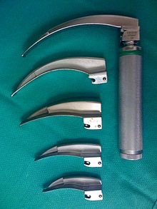
Laryngoscope handles with an assortment of Miller blades (large adult, small adult, child, infant and newborn) 
Laryngoscope handle with an assortment of Macintosh blades (large adult, small adult, child, infant and newborn) Tracheal tubes 
A cuffed endotracheal tube, constructed of polyvinyl chloride |
|
INDICATIONS FOR
ENDOTRACHEAL INTUBATION Inadequate ventilation, whether due to sedation and neuromuscular paralysis in the operating room, an obstructed or compromised airway, altered mentation, loss of consciousness, or respiratory failure can lead to brain injury or death within minutes. It is, thus, of great importance to know how to evaluate and address a patient who may require ventilatory support. INDICATIONS FOR ENDOTRACHEAL INTUBATION Indications for ENDOTRACHEAL INTUBATION in the operating room include: * the need to deliver positive pressure ventilation * protection of the respiratory tract from aspiration of gastric contents * surgical procedures involving the head and neck or in non-supine positions that preclude manual airway support * almost all situations involving neuromuscular paralysis * surgical procedures involving the cranium, thorax, or abdomen * procedures that may involve intracranial hypertension Some non-operative indications are: * profound disturbance in consciousness with the inability to protect the airway * tracheobronchial toilet * severe pulmonary or multisystem injury associated with respiratory failure, such as sepsis, airway obstruction, hypoxemia, and hypercarbia Objective measures may also be used to help determine the need for intubation: * respiratory rate > 35 breaths per minute * vital capacity < 15 ml/kg in adults and 10 ml/kg in children * inability to generate a negative inspiratory force of 20 mm Hg * PaO2 (arterial partial pressure of oxygen) < 70n mm Hg * A-a gradient (Alveolar-arterial) > 350 mm Hg on 100% oxygen * PaCO2 (arterial partial pressure of carbon dioxide) > 55 m Hg (except in chronic retainers) * dead space > 0.6 L HOW TO INTUBATE Prior to intubation, always check equipment and make sure everything you might need is not only within your reach, but also properly working. If in the operating room, a complete check of the anesthesia equipment at the start of each day as well as a modified check before each new case is imperative. If in the emergency room or the hospital wards, make sure you know where all of your equipment is and, also, that you have the necessary resources to support the patient once intubated. Prior to positioning the patient: * endotracheal tube Make sure that your laryngoscope is locked into position and that the incandescent light on the blade tip functions. Also make sure that you have several alternate blades available in case the one you have chosen does not allow for visualization of the cords. * Examine the endotracheal tube. Make sure that the cuff inflates by using a 10-mL syringe to inflate the cuff and then detach the syringe to ensure that the cuff pressure is maintained. Be sure to deflate deflate the cuff after testing it. * Attach the connector to the proximal end of the tube. Push it in as far as possible to lessen the likelihood of disconnection. * If you are going to use a stylet, it should be inserted into the ET tube and bent to resemble a endotracheal tube with stylethockey stick to facilitate intubation of an anteriorly positioned larynx. Even if you do not plan on using a stylet, one should be within easy access in case the intubation proves to be more difficult than anticipated. * Ensure a functioning suction unit to clear the airway in case of unexpected blood, emesis or secretions. * Ensure that you have tape within your reach to secure the tube once it is in place. Proper patient positioning can be the difference between a successful and failed intubation. * The patient’s head should be level with the physician’s xiphoid process. * To achieve the sniff position (which allows for optimal visualization of the glottic opening), elevate the patient’s head and extend the atlanto-occipital joint. This can be achieved by sliding your free hand (right hand if you are right handed, left hand if you are left handed) beneath the patient’s head and gently lifting it up and towards you. Or, you can gently position the chin up and mouth open before attemting laryngoscopy. * The "scissor technique" can also be used to further open the patients mouth. Cross your right forefinger and thumb and insert into the right side of the patient's mouth. Apply pressure to the upper teeth with your forefinger and the lower teeth with your thumb to open the mouth. Be sure to position your hands so as NOT to obstruct your view. Mask Ventilation is often used in the operating room after induction, prior to intubation. If you are able to achieve signs of ventilation using this technique, you are afforded the knowledge that, if intubation fails, you are able to achieve ventilation using the bag-mask-valve device. Further, it allows for pre-oxygenation. Preparation for induction and intubation in the operating room also involves pre-oxygenation with several (eight) deep breaths of 100% oxygen. Preoxygenation provides an extra margin of safety in case the patient is not easily ventilated after induction. After preoxygenating the patient and positioning the patient in the Sniff position, with the patient’s mouth widely open, carefully introduce the blade, held in your LEFT HAND, into the right side of the mouth. Regardless of which blade is used, IT MUST NEVER PRESS AGAINST THE TEETH or dental trauma will result. The tongue is then swept to the left and up into the floor of the pharynx by the blade’s flange. Macintosh BladesThe curved Macintosh blade is inserted past the tongue into the vallecula (at the base of the tongue). Providing sufficient lifting force in parallel with the handle, yet avoiding posterior rotation that causes the blade to press against the teeth, pressure is applied deep in the vallecular space by the tip of the blade immediately anterior to the epiglottis, which flips out of the visual field to expose the laryngeal opening. Miller Blade The straight Miller blade is inserted deep into the oropharynx, PAST the epiglottis. Providing sufficient lifting force in parallel with the handle, yet avoiding posterior rotation that causes the blade to press against the teeth, under direct vision, the blade is slowly withdrawn. It will slip over the anterior larynx and come to a position at which it holds the epiglottis flat against the tongue and anterior pharynx, exposing a view of the larynx. With either blade, the handle is raised up and away from the patient in a plane perpendicular to the patient’s mandible. Avoid trapping a lip between the teeth and the blade and AVOID using the teeth as leverage and avoid posterior rotation of the blade. Once a view of the larynx is obtained via laryngoscopy, the ETT is introduced with the RIGHT HAND through the right side often mouth. Directly observe the tip of the tube passing into the larynx, between the abducted cords. Pass the tube 1 cm through the cords. The ETT should lie in the upper trachea but beyond the larynx (3 to 4 cm proximal to the carina). If the patient is going to be repositioned, the cuff should be closer to 2 cm beyond the cords. Remove the laryngoscope, careful not to displace the ET tube and not to cause trauma to the teeth, lips or mucosa. Inflate the cuff with the least amount of air necessary to create a seal during positive pressure ventilation (usually 4-8 mL of air). Remove the mask from the bag-valve device and attach the 15 mm connector on the proximal end of the ET tube to the bag-valve device (into which oxygen is flowing and to which the carbon dioxide detector is attached). Provide positive pressure and immediately (and quickly): * ausculatate the chest for breath sounds * check the capnoraphic tracing on the monitor to ensure end tidal CO2 * check the connector for fog * look at the chest for expansion with each breath If there is any question as to whether the tube is in the esophagus or trachea, remove the tube, ventilate with a mask and try again, this time attempting to adjust anything that may have interfered with your first attempt. You might reposition the patient, use a different blade, decrease tube size, or add a stylet. If you are sure that your intubation is successful, turn on the mechanical ventilator. Continuously provide positive-pressure ventilation at a volume of 350-700 ML per 70 kg (5-10 mL/Kg) and at a sufficient rate to maintain normal end tidal CO2 (8-12 respirations per minute). Proceed to tape or tie the tube to secure its position. Do not tape or tie the cuff. To prevent the patient from biting and occluding the ETT during emergence from anesthesia, a roll of gauze can be placed between the teeth or an OPA can be inserted. Document the view of the larynx obtained during laryngoscopy using the following criteria: * Grade I: full view of the cords * Grade II: partial view of the cords * Grade III: view of the epiglottis * Grade IV: No view of the cords or epiglottis OTHER WAYS TO INTUBATE Nasotracheal Intubation: Nasal intubation is similar to oral intubation except that the ETT is advanced through the nose into the oropharynx before laryngoscopy. If the patient is awake, local anesthetic drops and nerve blocks can be used. A lubricated ETT is introduced along the floor of the nose, below the inferior nasal turbinate, perpendicular to the face. Often, a nasopharyngeal airway can be used. The tube is advanced until it can be visualized in the oropharynx. Via laryngoscopy, the tube is then advanced in between the abducted vocal cords. Nasal instrumentation (with ETTs, NPOs, or nasal catheters) is contraindicated in all patients with severe midfacial trauma. BougieBougie: The Bougie is a straight, semi-rigid stylette-like device with a bent tip that can be used when intubation is (or is predicted to be) difficult – often helpful when the tracheal opening is anterior to the visual field. During laryngoscopy, the bougie is carefully advanced into the larynx and through the cords until the tip enters a mainstem broncus. While maintaining the laryngoscope and Bougie in position, an assistant threads an ETT over the end of the bougie, into the larynx. Once the ETT is in place, the bougie is removed. Light Wand: Lightwands, when inserted into an endotracheal tube, may be useful for blind intubations of the trachea (when the laryngeal opening cannot be visualized). The end of the ET tube is at the entrance of the trachea when light is well transilluminated through the neck (the jack o’lantern effect). The tube can then be threaded off the light wand and into the trachea in a blind fashion. light wand Flexible Fiberoptic Bronchoscopy: Laryngoscopy may be contraindicated in a patient who requires intubation and mechanical ventilation. This is often the case in trauma patients who may have an unstable cervical spine or in patients with poor range of motion of the temporo-mandibular joint. In such patients, flexible fiberoptic bronchoscopy allows for indirect visualization of the larynx. The endoscope is introduced through the mouth or nose. Once anatomic structures are recognized, and the larynx or trachea are entered under direct visualization. COMPLICATIONS OF INTUBATION Complications of laryngoscopy and intubation are most frequently secondary to airway trauma, tube malpositioning, tube malfunction or physiologic responses to airway instrumentation. Trauma such as tooth damage, lip/tongue/mucosal laceration, sore throat, dislocated mandible, retropharyngeal dissection can occur during laryngoscopy and intubation. Mucosal inflammation and ulceration and excoriation of nose can occur while the tube is in place. Laryngeal malfunction and aspiration, glottic, subglottic or tracheal edema and stenosis, vocal cord granuloma or paralysis during extubation. Malpositioning of the endotracheal tube can result in esophageal intubation and unintentional extubation. Physiologic responses to intubation include hypertension, tachycardia, intracranial hypertension, and laryngospasm. Laryngospasm, which occurs during induction and recovery from anesthesia or, rarely, in an awake patient, is a forceful involuntary spasm of the laryngeal musculature caused by sensory stimulation of the superior laryngeal nerve. Treatment includes positive pressure ventilation via a bag-mask device using 100% oxygen or administration of IV lidocaine. HOW TO EXTUBATE Knowing when to extubate is also an important knowledge set. In general, it is best to extubate when a patient is still deeply anesthetized (but with adequate spontaneous respirations) or when the patient is awake and responsive with stable vital signs, good grip and sustained head lift. Adequate reversal of neuromuscular blockade must be established. A patient must also demonstrate adequate spontaneous respiratory function with a vital capacity of greater than 15 mL/kg and a negative inspiratory force of greater than 20 mm Hg. Extubation while the patient is in a light plane of anesthesia or still emerging from anesthesia is avoided because of an increased risk of laryngospasm, the most dreaded complication of extubation. Regardless of whether a patient is extubated while deeply anesthetized or awake, begin by thoroughly suctioning the patient’s pharynx and mouth in order to decrease the risk of aspiration or laryngospasm. Also, “preoxygenate” the patient with 100% oxygen in case it becomes difficult to establish an airway after the ETT is removed. Untape the ETT and deflate its cuff. Apply a small degree of positive pressure on the air bag to help blow out any secretions you may have missed on first suctioning and suction again. Withdraw the tube on end-inspiration or end-expiration in a single, smooth motion. Apply a face mask to deliver 100% oxygen. |
